Autophagy
Cellular pathways that deliver cytoplasmic material to lysosomes or vacuoles for degradation, quality control and recycling.
- Revision
- 1
- Created by
- SCIENDIA Knowledge Desk
- Updated by
- SCIENDIA Knowledge Desk
- Last updated
- 18.08.2026 12:20
Built by the community
Members can improve this article. Every saved change remains visible in the revision ledger.
Overview
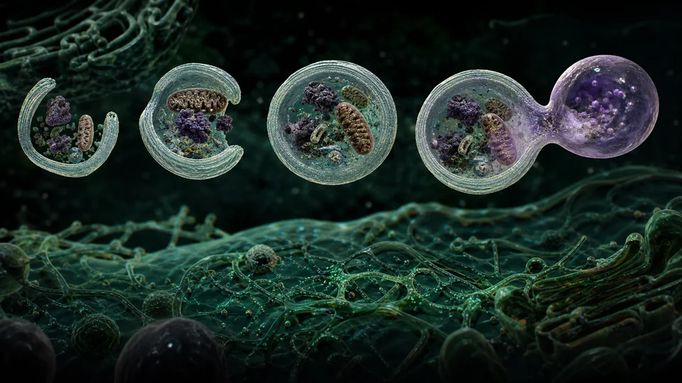
Macroautophagy encloses proteins, organelles or invading material in a double-membrane autophagosome that later fuses with a degradative compartment. Basal turnover protects cell quality, while nutrient stress increases recycling of macromolecular building blocks.
Technical foundations
Macroautophagy begins when nutrient and energy sensors regulate the ULK kinase complex and phosphatidylinositol-three-kinase machinery. A phagophore nucleates, expands through ATG conjugation systems and recruits LC3-family proteins. Cargo receptors such as p62 bind ubiquitinated targets and LC3, enabling selective removal of aggregates, mitochondria or pathogens. Closure creates a double-membrane autophagosome that fuses with lysosomes; acid hydrolases degrade cargo and transporters return amino acids, lipids and sugars to metabolism.
How it works
Nutrient and stress sensors regulate initiation complexes that nucleate an isolation membrane. Conjugation systems expand and close it, cargo receptors link selected targets to membrane proteins and lysosomal enzymes break contents into reusable components.
Measurement and research methods
Autophagic flux is measured by comparing LC3 turnover and cargo degradation with and without lysosomal inhibition, not by static vesicle number alone. Fluorescent tandem reporters distinguish acidic autolysosomes from earlier compartments, and electron microscopy confirms membrane ultrastructure. Genetic deletion of core ATG genes tests dependence but can have autophagy-independent effects. Tissue studies require timing, cell identity and nutrient state because basal flux varies. Controls distinguish increased formation from failed fusion or lysosomal dysfunction.
Key ideas
- More autophagosomes can indicate increased formation or blocked degradation.
- Bulk and selective autophagy serve different physiological roles.
- Autophagy can suppress early damage yet support survival of established disease cells.
Current research frontier
Research targets selective autophagy in neurodegeneration, infection, metabolism and cancer. Mitophagy maintains mitochondrial quality, xenophagy captures microbes and secretory autophagy routes selected cargo outside cells. Pharmacological activation might clear toxic proteins, whereas inhibition can sensitise some tumours; systemic effects make direction context-dependent. Open questions include membrane sources, receptor redundancy and age-related lysosomal decline. Clinical translation needs target-engagement biomarkers showing flux in the relevant tissue rather than assuming that blood markers reflect brain or tumour autophagy. Longitudinal human studies must also distinguish adaptive short-term recycling from chronic pathway failure and determine whether intervention improves tissue function rather than changing one molecular marker.
Why it matters
Autophagy connects metabolism, immunity, ageing, neurodegeneration, infection and cancer. Measuring its dynamic flux informs drug development and cell-stress biology.
Limits and open questions
Static markers are easily misinterpreted, and systemic inhibition affects many tissues. Cargo selectivity, membrane origin and stage-specific effects in human disease remain incompletely resolved.
Explore through connected concepts
This article is indexed with 20 technical tags. Select a tag to explore the Wiki by concept.
You may also explore
Recommended from the technical tags shared with this article.
Immunometabolism
The reciprocal control between cellular metabolic pathways and the activation, differentiation and function of immune cells.
Read article →