Bioprinting
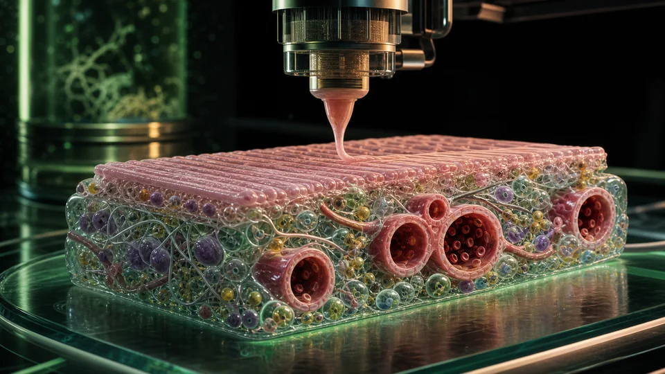
Layer-by-layer fabrication of living tissue constructs using cells, biomaterials and spatially controlled deposition.
- Revision
- 1
- Created by
- SCIENDIA Knowledge Desk
- Updated by
- SCIENDIA Knowledge Desk
- Last updated
- 18.08.2026 12:20
Built by the community
Members can improve this article. Every saved change remains visible in the revision ledger.
Overview
Bioprinting places cell-laden bioinks and supporting materials into designed three-dimensional architectures. The objective is not merely a shaped gel but a viable construct whose cells mature, communicate and develop mechanical and transport functions.
Technical foundations
Bioinks combine cells with hydrogels based on collagen, gelatin, alginate, fibrin or synthetic polymers. Rheology determines whether material flows through a nozzle yet retains shape after deposition; shear can damage cells. Ionic, thermal, enzymatic or light crosslinking stabilises constructs, but crosslink density affects diffusion, mechanics and cell migration. Extrusion handles viscous materials, inkjet methods place droplets and light-based printing polymerises entire patterns rapidly. Sacrificial inks create channels later removed for perfusion.
How it works
Extrusion, droplet or light-based systems deposit or solidify bioinks. Crosslinking stabilises geometry while pores and channels support diffusion or perfusion. After printing, biochemical and mechanical cues guide cell organisation, and bioreactors provide nutrients, flow or cyclic loading.
Measurement and research methods
Construct evaluation includes dimensional fidelity, cell viability, phenotype, mechanical properties and functional outputs over time. Live-dead staining immediately after printing does not prove maturation. Perfusion tests quantify channel integrity and transport, while histology, single-cell profiling and electrophysiology assess organisation. Independent print runs and cell donors capture process variation. Sterility, raw-material traceability and closed-system manufacture become essential for implantation. Models predict oxygen and nutrient gradients and help set maximum spacing between perfusable channels.
Key ideas
- Printability, cell viability and long-term tissue function are separate requirements.
- Vascular transport limits the size of living constructs.
- A digital geometry does not guarantee biological self-organisation after printing.
Current research frontier
Research focuses on prevascular networks that connect with host circulation, innervation, immune compatibility and organ-specific extracellular matrix. Volumetric printing speeds fabrication and organoid printing combines self-organisation with imposed geometry. Patient-derived cells enable personalised models, while universal cell sources require immune engineering. Open challenges include capillary-scale resolution over clinically relevant volume, long-term mechanical durability and reproducible differentiation. Regulatory strategies must define product identity when living constructs continue changing after manufacture, and therapeutic claims require comparison with simpler scaffolds or cell delivery.
Why it matters
Bioprinting supports disease models, drug testing, regenerative implants and research on tissue development. It can place multiple cell types more deliberately than spontaneous organoid culture.
Limits and open questions
Bioinks often trade mechanical strength against cell compatibility, and maturation can take weeks. Vascular integration, innervation, immune response, manufacturing reproducibility and regulatory classification remain major barriers to therapeutic-scale organs.
Explore through connected concepts
This article is indexed with 20 technical tags. Select a tag to explore the Wiki by concept.
You may also explore
Recommended from the technical tags shared with this article.
Organoids
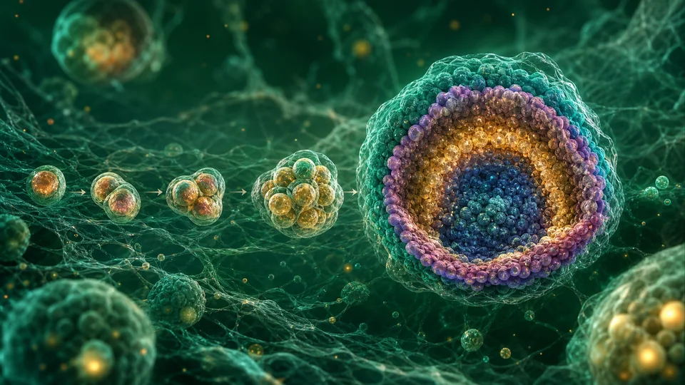
Three-dimensional, self-organising cell cultures that reproduce selected structural and functional features of organs.
Read article →