Liquid Biopsy
Analysis of tumour-derived DNA, cells, vesicles or other biomarkers in blood and body fluids to characterise cancer non-invasively.
- Revision
- 1
- Created by
- SCIENDIA Knowledge Desk
- Updated by
- SCIENDIA Knowledge Desk
- Last updated
- 19.08.2026 09:05
Built by the community
Members can improve this article. Every saved change remains visible in the revision ledger.
Overview
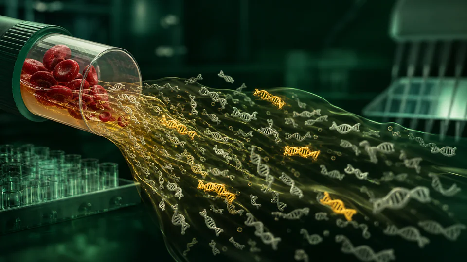
Liquid biopsy samples molecular material shed by tumours into circulation or other accessible fluids. Circulating tumour DNA can reveal somatic mutations, copy-number changes, methylation patterns and fragment characteristics, while circulating tumour cells and extracellular vesicles provide cellular or RNA information. The fraction of tumour-derived material varies strongly with disease site, burden and treatment.
Technical foundations
Cell-free DNA consists mainly of short nucleosomal fragments released during cell death. Tumour-derived molecules may carry mutations, altered methylation, copy-number imbalance or distinctive fragment endpoints. The expected count of rare molecules follows sampling statistics: at very low fraction, drawing more sequencing reads cannot recover molecules that never entered the tube. Molecular barcodes group copies from one original fragment and consensus calling suppresses polymerase and sequencing errors. Biological background from normal tissues sets an additional limit beyond instrument accuracy.
How it works
Plasma is separated promptly to limit contamination from lysed blood cells. Assays then enrich targeted genomic regions or sequence broadly with molecular barcodes that distinguish original molecules from amplification errors. Bioinformatic pipelines suppress sequencing noise and variants arising from age-related blood-cell clones. Results may support therapy selection, treatment-response monitoring, residual-disease detection or early-detection research.
Measurement and research methods
Assay validation uses reference materials spanning variant type and allele fraction, negative donors and reproducibility across operators and reagent lots. Pre-analytical studies test collection tube, processing delay, storage and plasma input. Analytical performance reports limit of blank, detection and quantification rather than one sensitivity number. Clinical studies compare plasma results with tissue, imaging and outcomes in the intended population. Longitudinal minimal-residual-disease assays require predefined sampling times and blinded thresholds to avoid interpreting noise after outcomes are known.
Key ideas
- A negative test may reflect low shedding or insufficient sampling rather than absence of cancer.
- Clonal haematopoiesis can imitate tumour mutations unless matched blood-cell analysis or validated filtering is used.
- Analytical detection, clinical validity and demonstrated patient benefit are separate levels of evidence.
Current research frontier
Research combines sequence, methylation, fragmentomics, proteins and clinical variables to improve early detection and tissue-of-origin prediction. Personalised assays track mutations selected from a patient's tumour, while tumour-naive tests seek broader use without tissue. Open questions include how to manage indeterminate positives and which intervention improves survival after molecular relapse is detected earlier than imaging. Population screening demands very high specificity because false positives accumulate when prevalence is low, making prospective outcome trials and equitable follow-up pathways essential.
Why it matters
Repeated blood sampling can track tumour evolution more easily than repeated tissue biopsy and may capture DNA from several metastatic sites. Liquid biopsy already supports selected molecular treatment decisions and is being studied for recurrence surveillance and multi-cancer screening.
Limits and open questions
Very low variant fractions demand stringent contamination control and statistical thresholds. Tissue remains necessary for histology and many diagnoses. Screening can create false positives, uncertain tissue-of-origin results and overdiagnosis, so clinical utility requires prospective trials showing that testing leads to earlier effective treatment without disproportionate harm.
Explore through connected concepts
This article is indexed with 20 technical tags. Select a tag to explore the Wiki by concept.
You may also explore
Recommended from the technical tags shared with this article.
Cancer Immunotherapy
Treatments that redirect, strengthen or release immune responses to recognise and control malignant cells.
Read article →